Organizers
Miguel Concha, LEO-Lab, U-Chile
Steffen Härtel, SCIAN-Lab, U-Chile
Summary
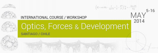
We cordially invite you to participate at the international course “Optics, Forces and Development”, to be held in Santiago, Chile, on May 5-16th 2014. The aim of the course is to train students, postdocs and young investigators from Latin America in theoretical and practical aspects of in vivo microscopy, and strategies for the visualisation and manipulation of cell and tissue morphodynamics in developing organisms.
The course is organised by the Biomedical Neuroscience Institute (Chile) and QuanTissue (ESF, Europe), and will coincide with the international symposium “Visualisation and manipulation of signals and forces in developing tissues” and the openlecture series on the “Origin of Animal Form in Evolution and Development”.
Location
Facultad de Medicina, Independencia 1027, U-Chile, Santiago
Central Topics:
- Principles of optics
- In vivo confocal imaging
- Image processing and analysis
- Force estimation in cells and tissues
- Laser micro-disecction
- Cytoskeletal dynamics
- Model Organisms (fish, fly, frog, mouse)
- Embryo manipulation (fish)
- Stem cells
Teachers, Students, and Program
Teachers, Students, and Program download here: PDF Document
Literature
- Super-Resolution Microscopy:
Nanoscopy by Using Blinking Enhanced Quantum Dots PDF Document
Superresolution imaging for neuroscience PDF Document
Widely accessible method for superresolution fluorescence imaging of living systems PDF Document - Forces and Microscopy:
Adhesion Functions in Cell Sorting by Mechanically Coupling the Cortices of Adhering Cells PDF Document
a-Catenin as a tension transducer that induces adherens junction development PDF Document
Deconstructing the Cadherin-Catenin-Actin Complex PDF Document - Differentiation of adult stem cells guided by matrix mechanics:
Cell responses to the mechanochemical microenvironment�Implications for regenerative medicine and drug delivery PDF Document
Hyaluronic acid matrices show matrix stiffness in 2D and 3D dictates cytoskeletal order and myosin-II phosphorylation within stem cells PDF Document
Matrix Elasticity Directs Stem Cell Lineage Specification PDF Document
Supplemental Literature: Cell locomotion and focal adhesions are regulated by substrate flexibility PDF Document - Image Processing:
Measurement of Instantaneous Velocity Vectors of Organelle Transport: Mitochondrial Transport and Bioenergetics in Hippocampal Neurons PDF Document
Reconstruction of Zebrafish Early Embryonic Development by Scanned Light Sheet Microscopy (supplementary material ) PDF Document PDF Document
3-D Active Meshes Fast Discrete Deformable Models for Cell Tracking in 3-D Time-Lapse Microscopy.pdf PDF Document
Manuals & More
ZEISS Principles of Confocal Microscopy PDF Document
Principles of Fluorescence Spectroscopy PDF Document
LEICE TCS LSI Brochure PDF Document
Huygens Professional User Guide from SVI: PDF Document Web Site
Intracellular Fluorescent Probe Concentrations by Confocal Microscopy, Finck et al. 1998 PDF Document
Seeing is believing? Alison J. North, The Journal of Cell Biology, Vol. 172, No. 1, January 2, 2006 9�18 PDF Document
The Good, the Bad and the Ugly ! Helen Pearson, NATURE, 447, May 2007 PDF Document
Measuring and interpreting point spread functions to determine confocal microscope resolution and ensure quality control, Cole et al 2011, NATURE PROTOCOL PDF Document
IDL/ScianTimeCalc: 1er Manual de Reconstrución 3D Word Document
Clases
Day 1
Clase: Steffen Härtel PDF Document
Clase Miguel Concha
Clase Martin Behrndt Zip Document
Day 2
Clase 5: Ulrich Kubitscheck A PDF Document
Day 3
Day 4
Clase: Jorge Jara A PDF Document
Clase: Jorge Jara BC PDF Document
Day 5
Clase: Ulrich Kubitscheck B PDF Document
Day 6
Clase: Antonio Jacinto PDF Document
Day 7
Clase: Roberto Mayor PDF Document
Clase: Xavier Trepat PDF Document
Day 9
Clase: Phillipp Keller PDF Document
Clase: Maria Elena Torres-Padilla PDF Document