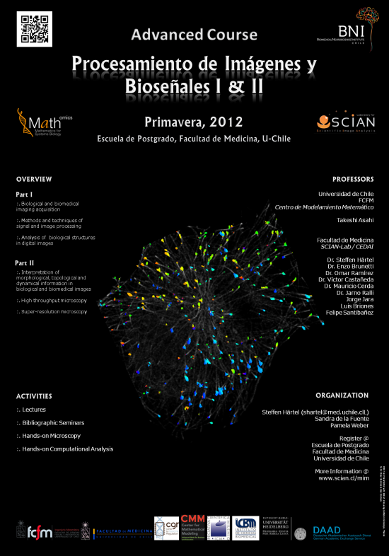
Santiago de Chile 18 de Agosto 2012 – 12 de Diciembre 2012, Facultad de Medicina, U-Chile
Organización
Steffen Härtel, SCIAN-Lab, BNI, ICBM, F-Med, Universidad de Chile
Localización
Facultad de Medicina, Independencia 1027, Universidad de Chile, Santiago, Chile.
Clases teóricas, prácticas y Seminarios:
Miércoles y Sábado| (ver programas): SCIAN-Lab
Exámenes:
17.10.2012| SCIAN-Lab
Profesores Participantes
ICBM | Facultad de Medicina, U-Chile
Dr. Steffen Härtel, SCIAN-Lab, Programa de Anatomía y Biología del Desarrollo (PABD)
Dr. Enzo Brunetti, Laboratorio Neuro-sistemas
Dr. Omar Ramírez, SCIAN-Lab, PABD
Dr. Víctor Castañeda, SCIAN-Lab, PABD
Dr. Mauricio Cerda, SCIAN-Lab, PABD
Dr. JarnoRalli, SCIAN-Lab, Universidad de Granada
Dr.(c) Jorge Jara, SCIAN-Lab, PABD
Ing. Luis Briones, SCIAN-Lab, PABD
Ing. Felipe Santibáñez, SCIAN-Lab
MSc. Susana Vargas, Centro de Espermiogramas Asistidos por Internet CEDAI-Spa, SCIAN-Lab
CMM | Facultad de Ciencias Físicas y Matemáticas (FCFM), U-Chile
Dr. Takeshi Asahi, Laboratorio de Modelamiento en Imágenes Científicas y Visualización(MOTIV) y Centro de Modelamiento Matemático (CMM)
Programa
El curso esta dividido en dos partes. Programa I y Programa II
Los tópicos centrales son:
(i) adquisición de imágenes biológicas y biomédicas,
(ii) métodos y técnicas de análisis y procesamiento de imágenes,
(iii) análisis de estructuras biológicas y biomedicas en imágenes digitales, y
(iv) microscopía de alta velocidad y super resolución.
Créditos:
Créditos para el curso se otorgan por la Facultad de Medicina, Universidad de Chile. Las materias podrán ser reconocidas para futuros estudios de postgrado, Diplomado y Magíster en Health Informatics.
Clases Bioseñales I
Sa 18.08 Clase 1: 3h 20min Steffen Härtel: Introduction, Microscopy, Fluorescence, Deconvolution. PDF File
Sa 18.08 Clase 2: 3h 20min Victor Casañeda: Detectores, X-Ray, NMR, MR, PET, SPECT, Ultrasound PDF File
Sa 25.08 Clase 3: 3h 20min Omar Ramirez / Steffen Härtel Praktico Microscopy (prepaso práctico 1-3).
Sa 25.08 Clase 4: 3h 20min Omar Ramirez / Steffen Härtel Praktico Microscopy, Flurescence, Deconvolution.
Mi 05.09 Clase 5: 3h 20min Takeshi Asahi: Introduction Fourier, Procesamiento Digital, Procesamiento Imágenes. PDF File , PDF File . PDF File
Mi 12.09 Clase 6: 3h 20min Enzo Brunetti: Teoría de señales: SEÑALES ELECTROFISIOLÓGICAS. PDF File
Mi 12.09 Clase 7: 3h 20min Jorge Jara: Métodos y técnicas de segmentación de imágenes I. PDF File
Sa 29.09 Clase 8: 3h 20min Jarno Ralli: Level Sets. PDF File
Sa 29.09 Clase 9: 3h 20min Jorge Jara / Luis Briones / Steffen Härtel Praktico Microscopy (prepaso práctico 4).
Mi 03.10 Clase 10: 3h 20min Mauricio Cerda: Análisis de estructuras biomédicas en imágenes digitales I. PDF File
Clases Bioseñales II
Mi 24.10 Clase 1: 3h 20min Jarno Ralli: Diffusion and Image Correspondences PDF File
Mi 07.11 Clase 2: 3h 20min Susana Vargas: Esperiogramas Digitales PDF File
Mi 14.11 Clase 3: 3h 20min Felipe Santibañez, Omar Ramirez: Resolución, Localisación y Colocalisación PDF File
Mi 28.11 Clase 4: 3h 20min Omar Ramirez, Victor Castañeda, Steffen Härtel: Super-Resolution Microscopy PDF File PDF File PDF File
Mi 05.12 Clase 5: 3h 20min Mallas Geométricas, Mauricio Cerda PDF File
Mayor Información
Contáctenos por email.
Preguntas Frecuentes
¿Cuanto cuesta el Curso?
- Si eres alumno regular de la Universidad de Chile, el curso es Gratuito.
- Si no eres alumno regular de la Universidad de Chile, el valor del curso es de 11,41 UF.
¿Donde me inscribo si “NO” soy alumno regular de post-grado de la Universidad de Chile?
- Si no eres alumno regular de post-grado de la Universidad de Chile, debes comunicarte con la oficina de post-grado de la facultad de Medicina de la Universidad de Chile, ademas debes descargar y enviar el formulario de solicitud de cupo para alumno libre desde AQUI .
- Mónica Astudillo, teléfono (56-2) 9786440, correo: mastudillo@med.uchile.cl
- María Isabel Peñaloza, teléfono (56-2) 9786713, correo: mpenaloza@med.uchile.cl
- Dirección Avenida Independencia 1027, Santiago, Chile.
¿Donde me inscribo si “SOY” alumno regular de post-grado de la Universidad de Chile?
- Para el caso de los alumnos de postgrado de otras facultades, cada secretaria de postgrado debe enviar la solicitud/inscripción de los alumnos que estén interesados en tomar el curso a la secretaria de postgrado de la facultad de medicina y deben incluir los siguientes datos (nombre, apellidos, programa/carrera, email, teléfono)
Grupos de Alumnos:
Grupo 1
Valentina Lagos (vlagosherrera@gmail.com)
German Fernández (gfernandez@uchile.cl)
Daniel Karmelic (DanielKarmelic@gmail.com)
Prepaso 4 (sa 29.09.2012) PDF File Excell File
Seminario 1 (mi 10.10.2012)
Grupo 2
Jorge Toledo (jorgetoledoh@gmail.com)
Daniel Droguett (ddroguett.o@gmail.com)
David Villaroel (davidbiovilla@gmail.com)
Prepaso 2 (sa 25.08.2012) PDF File
Seminario 5 ok (mi 10.10.2012)
Grupo 3
Vivina Torres (vatorresm@gmail.com)
Luis Baeza (luis.baeza@puc.cl)
Alejandra Lozano (juliaalejandra@gmail.com)
Prepaso 1 (sa 25.08.2012) PDF File
Seminario 4 (mi 10.10.2012)
Grupo 4
Juan José Ortega (juanjortega@gmail.com)
Carloz Núñez (nunezcarlos@live.cl)
Vera Solange (rivas.sole@gmail.com)
Prepaso 3 (sa 25.08.2012) PDF File
Seminario 3 (mi 10.10.2012)
Grupo 5
Verónica Bahamondes (bacoveronica@gmail.com)
Sebastián Fernández (sebferfrez@gmail.com)
Alex Córdova (cordova.alex@gmail.com)
Prepaso 5 (sa 06.10.2012) PDF File
Seminario 2 (mi 10.10.2012)
Prepasos Prácticos
- Bases de la Fluorescencia:
Principles of Fluorescence Spectroscopy (1st chapter) PDF Document
Fluorescent proteins: a cell biologist’s user guide. Erik Lee (2009), Trends in Cell Biology, Vol. 19(11) 649�655 PDF Document
Seeing is believing? Alison J. North, The Journal of Cell Biology, Vol. 172, No. 1, January 2, 2006 9�18 PDF Document - Bases de la Microscopía Confocal:
ZEISS Principles of Confocal Microscopy PDF Document
Live Cell Spinning Disk Microscopy. Graf et al. (2005) Adv Biochem Engin/Biotechnol 95:57-75 PDF Document
LEICE TCS LSI Brochure PDF Document
The Good, the Bad and the Ugly ! Helen Pearson, NATURE, 447, May 2007 PDF Document - Bases de la Deconvolución:
Huygens Professional User Guide from SVI: PDF Document
Intracellular Fluorescent Probe Concentrations by Confocal Microscopy, Finck et al. 1998 PDF Document - Histogramas (capítulo 3) y filtros basados en convolución (low-pass, detección de bordes, capítulos 2 y 4):
Feature Extraction and Image Processing, Nixon & Aguado (Elsevier) 2002. PDF Document - Momentos de morfología y descriptores de forma asociados:
Shape Analysis and Measument for HeLa cell classification of cultured cells in high throughput screening. Enamul Huqye Anm, Master thesis. (Leer sección 4: Methods) PDF Document
A Survey of Moment-Based techniques For unoccluded Object Representation and Recognition. Prokop et al. Graphical Model and Image Processing (1992), Vol 54 (5). (Leer secciones 1 y 2) PDF Document
Literatura para Seminarios
- In vivo Microscopy:
Cell tracking using a photoconvertible fluorescent protein. Hatta (2006) Nature Protocols PDF Document
Reconstruction of Zebrafish Early Embryonic Development by Scanned Light Sheet Microscopy. Keller (2008) Science 322:14 PDF Document
Escape Behavior Elicited by Single, Channelrhodopsin-2-Evoked Spikes in Zebrafish Somatosensory Neurons. Douglass (2008) Current Biology 18: 11:33 PDF Document - Localización y Colocalización:
Measurement of colocalization of objects in dual-color confocal images, Manders E. (1993) Journal of Microscopy 169: 375-382 PDF Document
A guided tour into subcellular colocalization analysis in light microscopy. Bolte S. et al (2006) Journal of Microscopy, 224 (3): 213�232 PDF Document
A guide to accurate fluorescence microscopy colocalization measurements. Comeau J.W., et al (2006) Biophys J. 91:4611-22 PDF Document
Accurate measurements of protein interactions in cells via improved spatial image cross-correlation spectroscopy. Comeau J.W.et al (2008) Mol Biosyst. 4: 672-85 PDF File
Multi-Image Colocalization and Its Statistical Significance. Fletcher P et al (2010) Biophys J. 99:1996-2005 PDF Document
Supporting Material: Multi-Image Colocalization and Its Statistical Significance. Fletcher P et al (2010) Biophys J. 99:1996-2005 PDF Document
Confined Displacement Algorithm Determines True and Random Colocalization in Fluorescence Microscopy. Ramirez O et al (2010) Journal of Microscopy, Sep 1;239(3):173-83 PDF Document
New Algorithm to Determine True Colocalization in Combination with Image Restoration and Time-Lapse Confocal Microscopy to Map Kinases in Mitochondria. Villalta et al (2011) PLOSone;6(4):e19031 PDF Document - Segmentación y aplicaciones:
A Methodology for Evaluation of Boundary Detection Algorithms on Medical Images. Chalana et al (1997) IEEE Transactions on Medical Imaging 16(5):642-652 PDF Document
Towards Objective Evaluation of Image segmentation Algorithms. Unnikrishnan et al (2007) IEEE Transactions on Pattern Analysis and Machine Intelligence 29(6):929-944 PDF Document
A framework for comparing different image segmentation methods and its use in studying equivalences between level set and fuzzy connectedness frameworks. Ciesielski et al (2011) Computer Vision and Image Understanding 115:721-734 PDF Document
Cell segmentation From 3-D Confocal Images of Early Zebrafish Embryogenensis. Zanella et al (2010) IEEE Transactions on Image Processing 19(3):770-781 PDF Document
3-D Quantification of the Aortic Arch Morphology in 3-D CTA Data for Endovascular Aortic Repair. Worz et al (2010) IEEE Transactions on Biomedical Engineering 57(10):2359-2368 PDF Document - Flujo Óptico y aplicaciones:
Performance of optical flow techniques for motion analysis of fluorescent point signals in confocal microscopy. Del Piano et al (2011) Machine Vision and Applications. PDF File
Measurement of Instantaneous Velocity Vectors of Organelle Transport: Mitochondrial Transport and Bioenergetics in Hippocampal Neurons. Akos et al (2008) Biophysical Journal 95:3079-3099 PPDF Document - Cuantificación topológica y aplicaciones:
Morphological analysis and modeling of neuronal dendrites. Van Pelt et al (2004) Mathematical Biosciences 188: 147�155 PPDF Document
Robust 3D reconstruction and identification of dendritic spines from optical microscopy imaging. Janoos (2009) Medical Image Analysis 13: 167 -179 PDF Document
Material Adicional
PDE Based Image Diffusion and AOS. Ralli (2012). PDF Document
