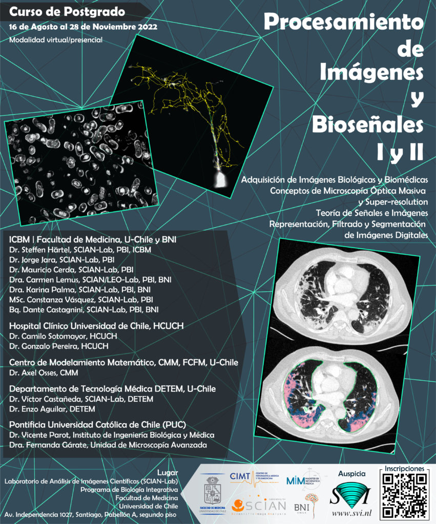
16 de Agosto – 28 de Noviembre 2022, SCIAN-Lab, Centro de Informática Médica y Telemedicina, Programa de Biología Integrativa, Facultad de Medicina, U-Chile
Organización:
Steffen Härtel / Jorge Jara-Wilde, SCIAN-Lab, PBI, ICBM, F-Med, Universidad de Chile.
Lugar:
SCIAN-Lab, Facultad de Medicina Campus Norte, U. de Chile, Av. Independencia 1027, Independencia, Santiago. Block A, Piso 2.
Profesores Participantes:
ICBM | Facultad de Medicina, U-Chile y BNI
Dr. Steffen Härtel, SCIAN-Lab, Programa de Biología Integrativa (PBI), ICBM
Dr. Jorge Jara, SCIAN-Lab, PBI
Dr. Mauricio Cerda, SCIAN-Lab, PBI
Dra. Carmen Lemus, SCIAN/LEO-Lab, PBI, BNI
Dra. Karina Palma, SCIAN-Lab, PBI, BNI
MSc. Constanza Vasquez, SCIAN-Lab, PBI
Bq. Dante Castagnini, SCIAN-Lab, PBI, BNI
Hospital Clínico Universidad de Chile, HCUCH
Dr. Camilo Sotomayor, HCUCH
Dr. Gonzalo Pereira, HCUCH
Centro de Modelamiento Matematico, CMM, FCFM, U-Chile
Dr. Axel Osses, CMM
Departamento de Tecnología Médica DETEM, Facultad de Medicina, U-Chile
Dr. Víctor Castañeda, SCIAN-Lab, DETEM
Dr. Enzo Aguilar, DETEM
Pontificia Universidad Católica de Chile (PUC)
Dr. Vicente Parot, Instituto de Ingeniería Biológica y Médica
Dra. Fernanda Gárate, Unidad de Microscopía Avanzada
Programa del curso
El módulo (M12) está dividido en dos cursos:
- Procesamiento de Imágenes y Bioseñales I, M12.1 con 4 créditos:
PDF Document - Procesamiento de Imágenes y Bioseñales II, M12.2 con 3 créditos:
PDF Document
Unidades:
Unidad 1: Adquisición de imágenes biológicas y biomédicas
Unidad 2: Conceptos de microscopía óptica masiva y super-resolution
Unidad 3: Teoría de señales e imágenes
Unidad 4: Representación, filtrado y segmentación de imágenes digitales
Clases Procesamiento de Imágenes y Bioseñales I
Martes 16.08, 18.00h, Sesión I: 3h 20min
S. Härtel: Introduccion al Curso I
PDF Document
S. Härtel: Adquisición de imágenes biológicas y biomédicas I
PDF Document
Jueves 18.08 18.00h, Sesión II: 3h 20min
S Härtel: Adquisición de imágenes biológicas y biomédicas II
PDF Document
Jueves 25.08 18.00h, Sesión I: 3h 20min
Adquisición de imagenes biológicas y biomédicas III
A. Osses: PDF Document
Jueves 01.09 18.00h, Sesión II: 3h 20min
S. Härtel: Adquisición de imágenes biológicas y biomédicas IV
PDF Document
J. Jara: Huygens Software y prepaso deconvolución
PDF Document
Martes 06.09 18.00h, Sesión I: 3h 20min
Conceptos de microscopía óptica masiva y super-resolution I
S. Härtel: PDF Document
V. Castañeda: PDF Document
Miércoles 14.09 18.00h, Sesión II: 3h 20min
Conceptos de microscopía óptica masiva y super-resolution II
S Härtel: PDF Document
V .Parot: PDF Document
Miércoles 21.09 18.00h, Sesión I: 3h 20min
V. Castañeda: Teoría de señales e imágenes I
PDF Document
Miércoles 28.09 18.00h, Sesión II: 3h 20min
V. Castañeda: Teoría de señales e imágenes II
PDF Document
Viernes 30.09 18.00h, Sesión I: 3h 20min
Teoría de señales e imágenes III
E. Aguilar: PDF Document
A.Osses: PDF Document
Lunes 03.10 18.00h, Sesión II: 3h 20min
J. Jara: Representación, filtrado y segmentación de imágenes digitales I
PDF Document
Miércoles 05.10 18.00h, Sesión II: 3h 20min
J. Jara: Representación, filtrado y segmentación de imágenes digitales II
PDF Document
Miércoles 12.10 18.00h, Sesión I: 3h 20min
J. Jara: Representación, filtrado y segmentación de imágenes digitales III
PDF Document
Clases Procesamiento de Imágenes y Bioseñales II
Lunes 17.10 18.00h, Sesión I: 3h 20min
J. Jara: Análisis de imágenes biológicas y biomédicas en series de tiempo I
PDF Document
Martes 18.10 18.00h, Sesión II: 3h 20min
M. Cerda: Análisis de imágenes biológicas y biomédicas en series de tiempo II
PDF Document
Viernes 21.10 18.00h, Sesión I: 3h 20min
M. Cerda: Análisis de imágenes biológicas y biomédicas en series de tiempo III
PDF Document
Lunes 24.10 18.00h, Sesión II: 3h 20min
M. Cerda: Interpretación de imágenes biológicas y biomédicas en series de tiempo IV
PDF Document
Miércoles 26.10 18.00h, Sesión I: 3h 20min
Aplicaciones en Laboratorios I
D. Castagnini: PDF Document
F. Gárate: PDF Document
Viernes 28.10 18.00h, Sesión II: 3h 20min
Aplicaciones en Laboratorios II
E. Aguilar: PDF Document
V. Castañeda: PDF Document
Jueves 03.11 18.00h, Sesión I: 3h 20min
Aplicaciones en Laboratorios III
C. Vásquez: PDF Document
Lunes 07.11 18.00h, Sesión II: 3h 20min
Sesión de Seminarios I
Lunes 14.11 18.00h, Sesión II: 3h 20min
Sesión de Seminarios II
Curso:
– Documentos y Literatura
1 Bases de la Fluorescencia
Principles of Fluorescence Spectroscopy (1st chapter) PDF Document
Fluorescent proteins: a cell biologist’s user guide. Erik Lee (2009), Trends in Cell Biology Vol. 19(11) 649-655 PDF Document
Seeing is believing? Alison J. North, The Journal of Cell Biology Vol. 172, No. 1 9-18, January 2, 2006 PDF Document
The Good, the Bad and the Ugly! Helen Pearson, NATURE 447, May 2007 PDF Document
Quantitative Imaging in Cell Biology (1st chapter) PDF Document
2 Bases de la Microscopía Confocal y Deconvolución
ZEISS Principles of Confocal Microscopy PDF Document
Live Cell Spinning Disk Microscopy. Graf et al. (2005) Adv Biochem Engin/Biotechnol 95:57-75 PDF Document
LEICE TCS LSI Brochure PDF Document
Huygens Professional User Guide from SVI: PDF Document Link Web
Intracellular Fluorescent Probe Concentrations by Confocal Microscopy, Finck et al. 1998 PDF Document
Quantitative Imaging in Cell Biology (chapters 5, 7, 9, 10) PDF Document
3 Segmentación
Feature Extraction and Image Processing, Nixon & Aguado (Elsevier) 2002. Histogramas (cap. 3) y filtros basados en convolución (pasa-bajos, detección de bordes, caps. 2, 4) PDF Document
Gold-standard and improved framework for sperm head segmentation. Chang et al. (2014). PDF Document
ACME: Automated Cell Morphology Extractor for Comprehensive Reconstruction of Cell Membranes. Mosaliganti et al. (2012). PDF Document
6 Descriptores de forma
Feature Extraction and Image Processing, Nixon & Aguado (Elsevier) 2002. Capitulo 7: Chain codes, basic and moments descriptors. PDF Document
Computational Methods for Analysis of Dynamic Events In Cell Migration. Current Molecular Medicine 14(2). Shape and topology section. PDF Document
Semi-automated quantification of filopodial dynamics. Constantino et al. (2008). PDF Document
Analysis of endoplasmic reticulum of tobacco cells using confocal microscopy. Radochova et al. (2005). PDF Document
– Literatura para Seminarios
1 High throughput in vivo microscopy and cell tracking:
Cell tracking using a photoconvertible fluorescent protein. Hatta (2006) Nature Protocols PDF Document
Reconstruction of Zebrafish Early Embryonic Development by Scanned Light Sheet Microscopy. Keller (2008) Science 322:14 PDF Document
2 Medical image Analysis:
Retrieving the intracellular topology from multi-scale protein mobility mapping in living cells. Baum (2014) Nature DOI: 10.1038/ncomms5494 PDF Document
Cell tracking using a photoconvertible fluorescent protein. Hatta (2006) Nature Protocols PDF Document
Reconstruction of Zebrafish Early Embryonic Development by Scanned Light Sheet Microscopy. Keller (2008) Science 322:14 PDF Document
Escape Behavior Elicited by Single, Channelrhodopsin-2-Evoked Spikes in Zebrafish Somatosensory Neurons. Douglass (2008) Current Biology 18: 11:33 PDF Document
3 Digital Pathology:
Cell tracking using a photoconvertible fluorescent protein. Hatta (2006) Nature Protocols PDF Document
Reconstruction of Zebrafish Early Embryonic Development by Scanned Light Sheet Microscopy. Keller (2008) Science 322:14 PDF Document
Escape Behavior Elicited by Single, Channelrhodopsin-2-Evoked Spikes in Zebrafish Somatosensory Neurons. Douglass (2008) Current Biology 18: 11:33 PDF Document
4 Localización y Colocalización:
Measurement of colocalization of objects in dual-color confocal images, Manders E. (1993) Journal of Microscopy 169: 375-382 PDF Document
A guided tour into subcellular colocalization analysis in light microscopy. Bolte S. et al (2006) Journal of Microscopy, 224 (3): 213 232 PDF Document
A guide to accurate fluorescence microscopy colocalization measurements. Comeau J.W., et al (2006) Biophys J. 91:4611-22 PDF Document
Accurate measurements of protein interactions in cells via improved spatial image cross-correlation spectroscopy. Comeau J.W.et al (2008) Mol Biosyst. 4: 672-85 PDF Document
Multi-Image Colocalization and Its Statistical Significance. Fletcher P et al (2010) Biophys J. 99:1996-2005 PDF Document
Supporting Material: Multi-Image Colocalization and Its Statistical Significance. Fletcher P et al (2010) Biophys J. 99:1996-2005 PDF Document
Confined Displacement Algorithm Determines True and Random Colocalization in Fluorescence Microscopy. Ramirez O et al (2010) Journal of Microscopy, Sep 1;239(3):173-83 PDF Document
New Algorithm to Determine True Colocalization in Combination with Image Restoration and Time-Lapse Confocal Microscopy to Map Kinases in Mitochondria. Villalta et al (2011) PLOSone;6(4):e19031 PDF Document
5 Segmentación y aplicaciones:
A Methodology for Evaluation of Boundary Detection Algorithms on Medical Images. Chalana et al (1997) IEEE Transactions on Medical Imaging 16(5):642-652 PDF Document
Towards Objective Evaluation of Image segmentation Algorithms. Unnikrishnan et al (2007) IEEE Transactions on Pattern Analysis and Machine Intelligence 29(6):929-944 PDF Document
A framework for comparing different image segmentation methods and its use in studying equivalences between level set and fuzzy connectedness frameworks. Ciesielski et al (2011) Computer Vision and Image Understanding 115:721-734 PDF Document
Cell segmentation From 3-D Confocal Images of Early Zebrafish Embryogenensis. Zanella et al (2010) IEEE Transactions on Image Processing 19(3):770-781 PDF Document
3-D Quantification of the Aortic Arch Morphology in 3-D CTA Data for Endovascular Aortic Repair. Worz et al (2010) IEEE Transactions on Biomedical Engineering 57(10):2359-2368 PDF Document
6 Flujo Óptico y aplicaciones:
An Implementation of Multiscale Combined Local-Global Optical Flow. Jara et al (2014) IPOL. PDF Document
Computation and Visualization of Three-Dimensional Soft Tissue Motion in the Orbit. Abramoff et al (2002) IEEE Transactions on Medical imaging 21(4). PDF Document
7 Cuantificación topológica y aplicaciones:
Spatial mapping and quantification of developmental branching morphogenesis. Short el al (2013) Development 140. PDF Document
Computing Multiscale Curve and Surface Skeletons of Genus 0 Shapes Using a Global Importance Measure. Reniers et al (2008) IEEE TRANSACTIONS ON VISUALIZATION AND COMPUTER GRAPHICS 14(2). PDF Document
8 Artificial Intelligence in Radiology:
Deep learning workflow in radiology: a primer. Insights Imaging 11, 22 (2020). https://doi.org/10.1186/s13244-019-0832-5 PDF Document
Artificial intelligence with deep learning in nuclear medicine and radiology. EJNMMI Phys 8, 81 (2021). https://doi.org/10.1186/s40658-021-00426-y PDF Document
9 Expansion Microscopy:
Expansion microscopy. Science (2015) DOI: 10.1126/science.1260088 PDF Document
Expansion microscopy with conventional antibodies and fluorescent proteins. Nature methods (2016) https://doi.org/10.1038/nmeth.3833 PDF Document Suplementary Material
Q&A: expansion microscopy. BMC biology (2017) https://doi.org/10.1186/s12915-017-0393-3 PDF Document
10 Review: Microscopy Diffraction Barrier:
Breaking the Diffraction Barrier: Super-Resolution Imaging of Cells. Huang B. et al (2010) Cell 143:1047-1058 PDF Document
10.1 STED-Microscopy:
STED-Microscopy: Concepts for nanoscale resolution in fluorescence microscopy. Hell S. et al (2004) Current Opinion in Neurobiology 4:599-609 PDF Document
Microscopy and its focal switch. Hell S. (2009) Nature Methods. 6(1):24-32 PDF Document
10.2 SIM-Microscopy:
Subdiffraction multicolor imaging of the nuclear periphery with 3D structured illumination microscopy. Schermelleh et al (2008) Science, 320(5881):1332-6 PDF Document
Three-dimensional resolution doubling in wide-field fluorescence microscopy by structured illumination. Gustafsson et al (2008) Biophys J, 94(12):4957-70 PDF Document
Nonlinear structured-illumination microscopy: Wide-field fluorescence imaging with theoretically unlimited resolution Gustafsson (2005) 1381242953.6801PNAS: 13081-13086 PDF Document
8.3 PALM/STORM-Microscopy:
Imaging intracellular fluorescent proteins at nanometer resolution. Betzig et al (2006) Science, 313(5793), 1642-5 PDF Document
Super-resolution imaging by nanoscale localization of photoswitchable fluorescent probes. Bates M et al (2008) Curr Opin Chem Biol, 12(5): 505-514 PDF Document
A New Approach to Fluorescence Microscopy. Bates M (2010) SCIENCE 330: 1334-5 PDF Document
Superresolution Imaging of Chemical Synapses in the Brain. Dani A et al (2010) Neuron 68, 843-856 PDF Document
